Observing a Plant’s Vascular System with X-Ray Video

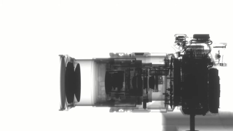
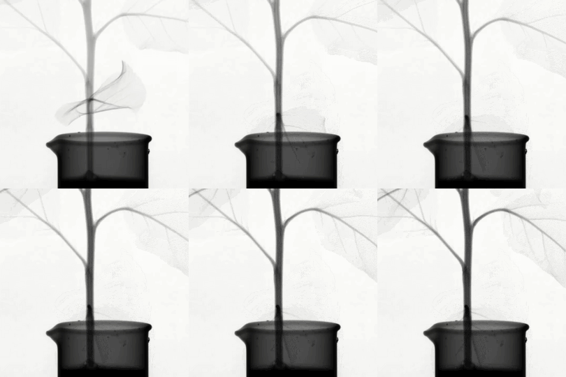
[Ben Krasnow] has a knack for showing us what’s inside of things while they’re moving. This week’s Applied Science experiment has him making time-lapse X-ray videos of things. This plant’s vascular system is just one of a few examples, the others being a dial clock and the zoom lens on a DSLR.
 The trick here is having an X-ray sensing panel that can be reused. It takes around five seconds of exposure to grab each 40×40 cm frame which are then assembled back into video.
Now watching mechanisms move is cool — [Ben’s] video back in 2015 to show what a phonograph needle in the groove of a vinyl record looks like under a scanning electron microscope is still one for the coolest “camera tricks” we’ve ever seen pulled off. But watching the vascular system of a plant function is the recipe for one of those ah-ha educational moments, so we hope that 7th-grade biology teachers everywhere will find their way to this video.
The apparatus is described in great detail, but regular Hackaday readers will most likely want to focus in on the teardown of the X-ray panel, which [Ben] describes as a giant digital camera sensor tuned for receiving the X-rays. The source is a 50 kV 1 mA tube that he compares to what is used at the dental office. (Obviously this requires forethought to ensure his automated time-lapse setup will fail safe with the X-ray tube.) A Cyclone III FPGA drives the panel, communicating with the sensor array via two Ethernet interfaces.
A friend sent a the broken panel to [Ben] and he was able to easily repair a MOSFET that got knocked out of place. [biluni] shows up in the comments of this video, sharing his recollection from working in the industry 15 years ago that a panel like this would have cost $150k! But considering the stellar resolution, and repeatable use, it sure as heck beats the old film process.
Post a Comment